June 2022
Clinical History
A female patient in her 20s presented to the emergency room with abdominal pain and hypertension was found to have a 10 cm exophytic mass at the superior pole of the right kidney on CT imaging which was read as a hemorrhagic cyst or complex cystic mass of possible adrenal origin. Follow up imaging revealed no other metastases. Adrenal workup with assessment of plasma free metanephrines, DHEA-S, 17-hydroxyprogesterone, testosterone, serum aldosterone to renin ratio and low-dose dexamethasone suppression test was significant for markedly elevated normetanephrine levels (30x the upper limit of normal), clinically suggesting paraganglioma versus pheochromocytoma. The patient was started on phenoxybenzamine (alpha blocker antihypertensive medication) to regulate the blood pressure and then right adrenalectomy was pursued for definitive management. Intraoperatively, the mass was difficult to dissect from critical structures and noted to have a cystic component. The specimen was submitted to pathology.
Gross findings
Specimen designated as right adrenal mass, weighed 170g and measured 12 x 8 x 4 cm. The specimen was soft, friable, hemorrhagic, and partially necrotic. Cut surfaces were variegated. A small portion of normal adrenal tissue was noted at the periphery.
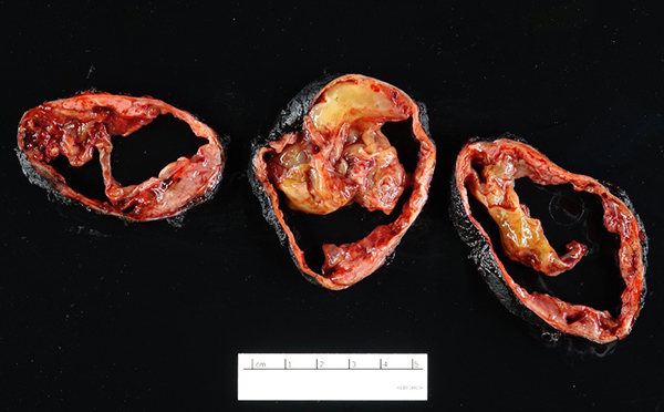
A. Sectioned mass with a fibrous capsule, revealing cystic degeneration and a heterogenous appearance.
Microscopic findings
Pheochromocytoma was identified comprising 50% of tumor volume. In addition, the lesion contained a primitive round cell component possibly neuroblastoma with focal neuroblastic differentiation (20%) and a malignant peripheral nerve sheath tumor (30%) with heterologous elements (cartilage). Areas of hemorrhage and necrosis were noted resulting in cystic degeneration.
Ancillary studies
Synaptophysin and chromogranin were positive in pheochromocytoma areas. S100 and neurofilament were focally positive in spindle cell areas and sustentacular cells. Vimentin was positive in spindle cell areas. H3K27me3 showed focal areas with loss of expression. Inhibin was positive in focal areas, consistent with entrapped cortical cells.
Diagnosis
Composite Pheochromocytoma with primitive round cell component (possibly neuroblastoma with focal ganglioneuroblastic differentiation) and a malignant peripheral nerve sheath tumor (MPNST) with heterologous elements.
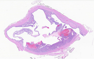
B. Complex cystic architecture (low power)
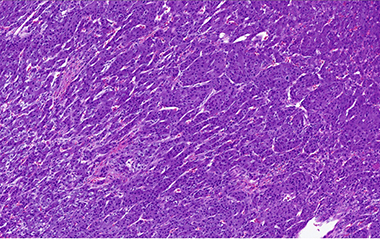
C. Pheochromocytoma with "zellballen" features
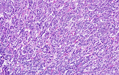
D. Pheochromocytoma
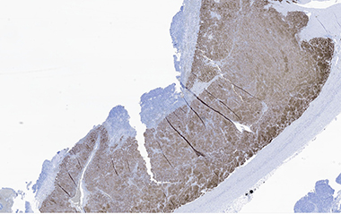
E. Pheochromocytoma with positive synaptophysin stain
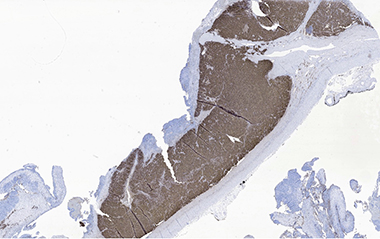
F. Pheochromocytoma with positive chromogranin stain
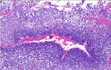
G. Primitive round cell component (high power)
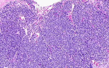
H. Primitive round cell component
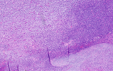
I. Malignant peripheral nerve sheath tumor with heterologous component
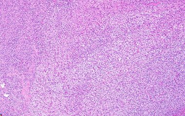
J. Malignant peripheral nerve sheath tumor
Discussion
Composite adrenal pheochromocytomas (CPs) are defined as pheochromocytoma with an adrenal medullary tumor, also of neural crest origin such as neuroblastoma (NB), ganglioneuroblastoma (GNB), ganglioneuroma (GN), benign nerve sheath tumor or malignant peripheral nerve sheath tumor (MPNST). CPs that include a MPNST component are extremely rare (less than 10 reported as of 2021). CPs are found with the following frequency of incidence: pheochromocytoma in combination with ganglioneuroma (75%), with ganglioneuroblastoma (10%), with neuroblastoma (10%) and with MPNST (most uncommon).
CPs are usually seen in adults 40-50 years in age with no gender predilection. Grossly, these tumors are expected to resemble usual pheochromocytoma and the mixed morphology are only detected under the microscope. We share this case because it is important to be aware of CPs and the extensive sampling that is necessary to detect them.
Differential diagnoses to consider include collision tumor, pure ganglioneuroma, ganglioneuroblastoma, or neuroblastoma, tumor-to-tumor metastasis and mix corticomedullary tumor.
Microscopically, transition between the tumors may be blended or abrupt, pheochromocytoma is almost mostly the predominant pattern with apparent zellballen or variant patterns, GN contains ganglion cells in background of Schwann cells, GNB will have immature and maturing neurons, neuropil and Schwann cells, NB will have small round blue cells and MPNST will show malignant spindle cells with high mitotic count. Note that to designate as composite, although there is no precise portion of the components specified as a definition, there must be complete tumor patterns and not just scattered neurons. Overall, patterns of growth, intermixed cellular components, and immunohistochemistry usually allow this tumors separation from its differential diagnoses.
The expected ancillary testing profile is pheochromocytoma showing diffuse chromogranin A and synaptophysin staining while ganglioneuroma, ganglioneuroblastoma and neuroblastoma shows weak focal staining mostly in processes. Schwann cells and sustentacular cells are expected to stain for S100.
30% of pheochromocytomas occur in a hereditary setting including NF1, MEN2A and 2B, and Von Hippel-Lindau (VHL) due to succinate dehydrogenase, RET and VHL genes. The most recent review of six CPs with MPNST shows one case with a history of NF1. Although CPs are mostly sporadic in nature, it is essential to do genetic studies or use surrogate immunohistochemical markers to rule out familial association.
Patient's young age should raise the question of von hippel-lindau associated versus succinate dehydrogenase (SDH) associated mutation. However, neither mutation was confirmed after molecular testing in our case.
Biologic behavior of pheochromocytoma is difficult to predict based on histology alone. Long-term follow up is required. Completely excised tumors do not usually recur or metastasize. They can be indolent even when ganglioneuroblastoma or neuroblastoma elements are present. Metastasis is rare and often comprise of the neural component. The prognosis of CP with MPNST is unknown, given the small number of reported cases. In our case, the patient was found to have recurrent metastasis in the abdomen within a short interval. The sampled material was small but contained the undifferentiated component which unfortunately predicted a poor outcome.
References
- Namekawa T, Utsumi T, Imamoto T, Kawamura K, Oide T, Tanaka T, Nihei N, Suzuki H, Nakatani Y, Ichikawa T. Composite pheochromocytoma with a malignant peripheral nerve sheath tumor: Case report and review of the literature. Asian J Surg. 2016 Jul;39(3):187-90.
- Mete O, Asa SL, Gill AJ, Kimura N, de Krijger RR, Tischler A. Overview of the 2022 WHO Classification of Paragangliomas and Pheochromocytomas. Endocr Pathol. 2022 Mar;33(1):90-114.
- Mukherjee S, Mondal A, Sengupta M, Chatterjee U, Sarkar D, Mukhopadhyay S. Composite phaeochromocytoma with malignant peripheral nerve sheath tumour: A case report with summary of prior published cases. Indian J Pathol Microbiol. 2021 Jul-Sep;64(3):571-574.
- Shida Y, Igawa T, Abe K, Hakariya T, Takehara K, Onita T, Sakai H. Composite pheochromocytoma of the adrenal gland: a case series. BMC Res Notes. 2015 Jun 24;8:257.