
June 2025 Case
Clinical History
Less than a week old male neonate was born presenting with a large pedunculated pigmented lesion on the forehead (3 cm in diameter), and up to a hundred or more well-defined pigmented papules (< 3 mm each) on the skin surfaces of the head, neck, trunk, extremities, perineum, and in the placenta. Abdominal ultrasound imaging demonstrated 7 hypoechoic hepatic foci (largest 5 mm), which is suggestive of possible liver involvement by this process on the skin versus a possibility of hemangiomas. The neonate had no evidence of lymphadenopathy or involvement of other organs including the central nervous system and lungs. The mother of this infant is healthy and without melanoma.
Histology
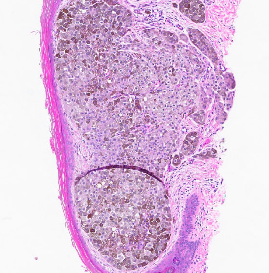
Figure 1: H&E stain at 10x magnification displaying atypical melanocytes proliferative within the dermis.
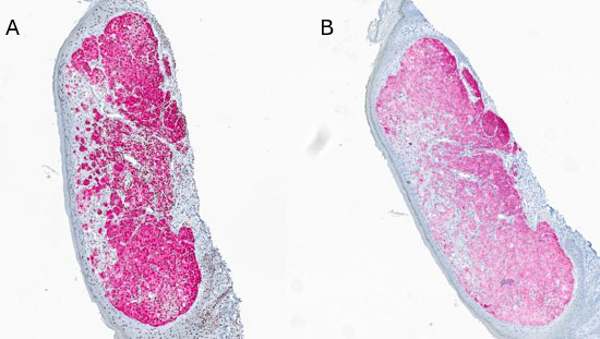
Figure 2: A - Melan A in red staining positive for the atypical melanocytes within the dermis. B - HMB 45 staining positive for the atypical melanocytes.
Diagnosis
Atypical congenital dermal melanocytic neoplasm with spitzoid features, ETV6:: NTRK3 gene fusion, and uncertain malignant potential, margins involved
Molecular Findings
Genomic profiling detected an ETV6::NTRK3 fusion and an NTRK3 amplification. The fusion was identified to occur between exons 4 of ETV6 fusing to exon 14 of NTRK3. The same ETV6::NTRK3 fusion was detected in the patient's mother's placenta as well.
Discussion
Dermatopathology consultation suggested the differential diagnosis include eruptive-disseminated atypical Spitz or spitzoid melanocytic neoplasms (EDSN) developing in utero with an ETV6:: NTRK3 fusion and uncertain malignant potential; and atypical spitzoid melanocytic neoplasms suspicious for melanoma with metastatic dissemination. Eruptive-disseminated Spitz nevi/tumors (EDSN) is an important stimulant of metastatic melanoma. Among all reported cases of EDSN, none of the patients have shown transformation to melanoma or died with follow-up as long as 26 years. This specimen failed to show sufficient high-risk morphological and genetic criteria for clear-cut malignancy. The placental involvement argues against a malignant process and potential liver involvement has not yet been pathologically confirmed. However, hypothetical liver metastases could still be considered within the spectrum of apparent benign metastases of this entity.
The ETV6::NTRK3 fusion has been detected in various tumor types including pediatric tumors such as childhood melanocytic tumors and infantile fibrosarcomas. Although this lesion did not fit the diagnostic criteria for Melanoma, one possible targeted therapy approach worth consideration is larotrectinib and entrectinib for solid tumors harboring an NTRK gene fusion. Larotrectinib is FDA-approved for adult and pediatric patients with solid tumors harboring an NTRK gene fusion.
In summary, this was not a straight forward diagnosis, and instead required the combination of expert histological, immunohistochemical, and molecular interpretation. This case brings attention to the importance of NTRK fusions in pediatrics patients. These lesions involve a broader panel of fusion partners and a wider range of tumors than previously realized. Furthermore this case provides an opportunity for a targeted therapy option in Latrectinib, which is a selective TRK inhibitor that works by blocking the activity of tropomyosin receptor kinases (TRK) encoded by the NTRK1, NTRK2, and NTRK3 genes. This case serves as support for the necessity of further research into histologic/molecular diagnostic discoveries, which will ultimately aid us in uncovering novel treatment strategies.
References
- Fernandez-Flores A, Cassarino D. Genetic Studies on a Case of Eruptive Disseminated Spitz Nevus and Review of Other 33 Cases. Am J Dermatopathol. 2022;44(12):989-1002. doi:10.1097/DAD.0000000000002310
- Wang L, Busam KJ, Benayed R, et al. Identification of NTRK3 Fusions in Childhood Melanocytic Neoplasms. J Mol Diagn. 2017;19(3):387-396. doi:10.1016/j.jmoldx.2016.11.005
- Kang J, Park JW, Won JK, et al. Clinicopathological findings of pediatric NTRK fusion mesenchymal tumors. Diagn Pathol. 2020;15(1):114. doi:10.1186/s13000-020-01031-w
- Zhao X, Kotch C, Fox E, et al. NTRK Fusions Identified in Pediatric Tumors: The Frequency, Fusion Partners, and Clinical Outcome. JCO Precis Oncol. 2021;1:PO.20.00250. doi:10.1200/PO.20.00250